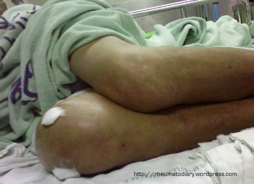
A 60 years old woman had been suffering from chronic pain and swelling of the right shoulder for two years. She had no fever and no history of weight loss. She had no prior medical problems. The examination revealed markedly swollen right shoulder, limited range of motion and mild tenderness. Synovial biopsy was done and Mycobacterium tuberculosis was found in the culture. Tuberculous arthritis of right shoulder was diagnosed. To my horror, she had never had a chest x-ray despite her frequent visit to the Orthopedics OPD! She was later treated with oral anti-TB drugs.
Case 3
July 23, 2010
Gout, Septic Arthritis Gout, Septic arthritis Leave a comment
This middle-aged man was admitted in the general wards because he had high fever and right knee pain for two days. As the photo shows, his right knee was swollen and warm and severely tender on examination. Joint aspiration was done. The synovial fluid exam revealed septic ranged WBC counts, Monosodium urate crystal was found from light microscopic exam and fluid gram stain showed gram-positive cocci bacteria. The diagnosis of septic arthritis and acute gouty arthritis was made. He was treated with intravenous antibiotics for the infection and intramuscular ACTH for his Gouty arthritis.
Case 2
June 19, 2010
CPPD, Osteoarthritis CPPD, Osteoarthritis Leave a comment
I did a case presentation last week. The topic was Osteoarthritis. So I chose one of the patients who came to the OPD for regular followup. She was 77 years old and obese. She was diagnosed with Osteoarthritis of both knees for two years. The examination revealed varus deformity (very obvious when she was standing – see photo), knee crepitus on motion and laxity of anterior cruciate ligaments.
I also reviewed her knee plain radiographs which were done 2 months ago but no one seemed to care about. To my surprise, the films not only displayed typical features of OA knee (joint space narrowing in medial compartment, subchondral bone sclerosis and osteophyte) but also showed chondrocalcinosis (abnormal calcification) in the joint space. Plus, on lateral view, narrowing of patello-femoral joint, scleroses of patella and bone formation at the superior pole of patella (Patella-wrapping signs) were also observed.
In short, she probably had CPPD (calcium pyrophosphate dehydrate deposition) as well despite the lack of history of acute knee inflammation.
Case 1 (part 2)
February 28, 2009
Non-rheumatic diseases, Systemic Lupus Erythematosus Peutz Jegher syndrome, SLE Leave a comment
This is the continuation of the previous post about a middle aged woman who suffered from infected ovarian endometrioma, beta-Thalassemia/HbE disease and SLE. During her admission, someone noticed abnormal lesion in her mouth.
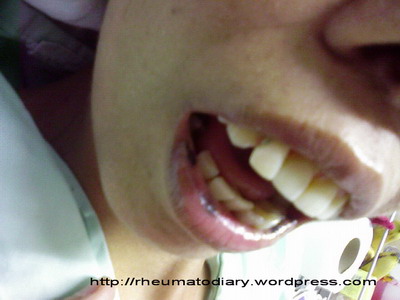
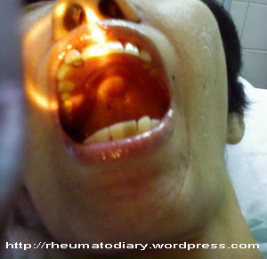
The physical examination revealed multiple hyperpigmented lesions, approximately 2-5 mm. in diameter with sharp border at her upper and lower lips, buccal mucosa and hard palate. There were also a few small lesion at her finger tips of both hands (I can’t believe I missed them when I examined her hand earlier.). We consulted the Dermatologist and he suspected the lesions could be the sign of Peutz Jegher Syndrome although she had no family history of any gastrointestinal polyps. The stool occult blood was negative. The patient was then scheduled for Barium enema and GI follow through to screen for polyposis. I am very sorry that I won’t be able to learn the results since my rotation ended here. If the patient really had PJS, It would be really absurd. No one should suffer from three major diseases at once (Thalassemia major, SLE and PJS).
p.s. the photoes are not very clear because I had no proper flash light when I took them.
Case 1
February 21, 2009
Systemic Lupus Erythematosus SLE 1 Comment
I met this patient during my last few weeks before graduation. A 42 year old woman came to OB-GYN emergency department because she had severe lower abdominal pain and fever for a day. She had been diagnosed for beta-thalassemia/Hb E disease since childhood. The physical examination showed high fever, marked pallor, marked tenderness and guarding in lower abdomen. Per vaginal examination and bedside ultrasonography lead to provisional diagnosis of tubo-ovarian abscess or infected endometrioma. She went through emergency explore laparotomy to TAH and BSO that night and the post operative diagnosis was infected endometrioma.
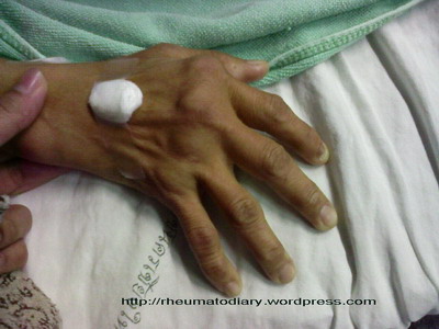
I first noticed the deformity of her hands just before she went to the operative room. When I asked about her hands, she told me she had been suffering from chronic pain and swelling of hand, wrist and knee joints for almost 2 years but she had no morning stiffness. No definitive diagnosis was established. On examination, she had ulnar deviation and muscle atrophy of both hands. Swelling with boggy consistency and mild tenderness of both PIP, MCP and wrist joints were found. I was pretty sure she had Rheumatoid arthritis.
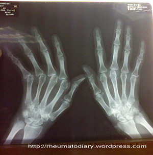
After the operation and her infection was well controlled, she was evaluated by a Rheumatologist. Her serologic test for Rheumatoid factor came up negative and plain radiograph of her hands showed subluxation of 2-5th MCP joints with no bony erosion. Further tests were done; her ANA was positive with 1280 titer and homogenous pattern, her anti-dsDNA was negative, her complete blood count showed lymphopenia. Other tests were pending. It’s a shame I could not follow her progression but at the time I finished my rotation, her diagnosis was possible SLE. The deformity of her hand was called Jaccoud Deformity.
I guess that’s about it for today.


